


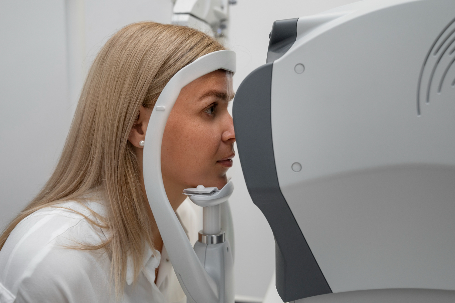
Basic Optical Coherence Tomography (OCT) interpretation programme
Optical Coherence Tomography (OCT) has become an essential tool in everyday ophthalmic practice, providing detailed, non-invasive images of the retina and optic nerve. As OCT use continues to expand across eye care settings, the ability to confidently and safely interpret scans is an increasingly important skill for the wider ophthalmic workforce.
This one-day, interactive study day is designed to give ophthalmic healthcare professionals a strong foundation in OCT principles and their clinical application. Through a mix of taught sessions and hands-on workshop activity, participants will build the knowledge and confidence to interpret normal scans, recognise common pathologies, and know when findings need to be escalated.
Course aims
To provide ophthalmic healthcare professionals with a strong foundation in OCT principles and their clinical application, enabling the safe interpretation of normal scans and the recognition of common ocular pathologies in everyday practice.
Learning outcomes
By the end of this course, participants will be able to:
- Explain OCT principles – describe the fundamental physics, image acquisition process, and core components of OCT technology.
- Identify normal OCT anatomy – recognise and label key retinal and optic nerve structures on standard OCT scans.
- Differentiate normal vs abnormal scans – distinguish normal anatomical variations from early or common pathological changes.
- Interpret common ocular pathologies – identify OCT features associated with frequently encountered conditions such as AMD, diabetic macular oedema, vitreomacular interface disorders, and glaucoma.
- Apply OCT findings to clinical decision-making – use OCT interpretation to support safe clinical judgement, appropriate escalation, and timely referral.
- Evaluate scan quality – assess image quality, recognise common artefacts, and understand how they may affect interpretation.
- Communicate OCT findings effectively – summarise OCT results clearly and accurately when discussing cases with colleagues or escalating concerns.
Course structure
Delivered across one full day, the programme moves from core theory through to applied, case-based practice:
- Foundations – retinal and optic nerve anatomy, and an introduction to how OCT works and its clinical applications.
- The normal scan – key features of a healthy retina and how to read an OCT report.
- Common pathologies – OCT features of AMD, diabetic retinopathy, glaucoma, central serous retinopathy, and vitreoretinal conditions.
- Practical application – recognising when OCT is essential, understanding its constraints, and communication and escalation.
- Scan quality and artefacts – differentiating normal from abnormal findings and identifying poor-quality scans.
- Interactive workshop – small-group interpretation of sample OCT scans, followed by an open Q&A.
Programme director
Anjali Praveen, Head of Nurse Education, Moorfields Eye Hospital NHS Foundation Trust.
Who is this programme for?
Ideal for those who are beginning to use OCT in clinical practice and who want to build confidence in scan interpretation, image quality assessment, and recognition of common pathologies, including:
- Ophthalmic nurses
- Orthoptists
- Healthcare support staff/Technicians
Book Now
| Date | Location | Time | Seats | Price | |
| Saturday 24 April 2027 | Education Hub, 15 Ebenezer Street, London N1 7NH | 22 | £350.00 | ||
| Saturday 24 April 2027 | Online via MS Teams | £275.00 |